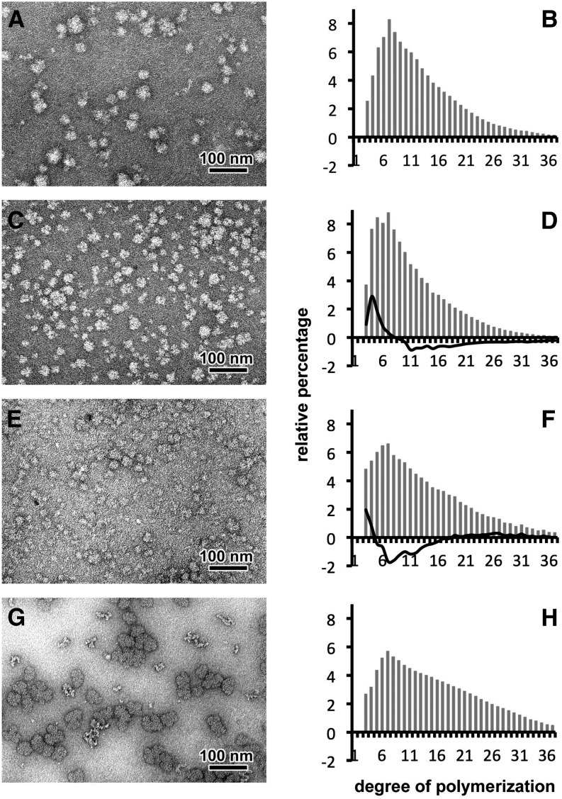
Figure 2.
Characterization of WSPs Accumulated in the Wild-Type Strain and Class A Mutants of Cyanobacterium sp CLg1.
Negative staining following TEM observations suggest that WSP of the wild type (A), the 99A7 mutant (C), and the 153H12 mutant (E) are highly branched polysaccharides with a diameter below 50 nm similar to glycogen particles of rabbit liver (G). After purification and complete digestion with a commercial isoamylase, chains of Glc were separated according to their DP by HPAEC-PAD. From three independent extractions, the relative abundance for each DP (gray bars) was determined for the wild-type strain (B), the severe mutant 99A7 (D), the intermediate 153H12 mutant (F), and glycogen from rabbit liver (H). Subtractive analyses (percentage of each DP of mutant minus percentage of each DP in the wild type), depicted as black lines in (D) and (F), reveal an increase of short chains (DP 3 to 7) and a defect in long-chain content (DP 12 to 35) in the WSPs accumulated in the mutant 99A7 (in [D]) and in the intermediate mutant 153H12 (in [F]). WSP appears to contain fewer chains with a DP ranging between 4 and 16.

