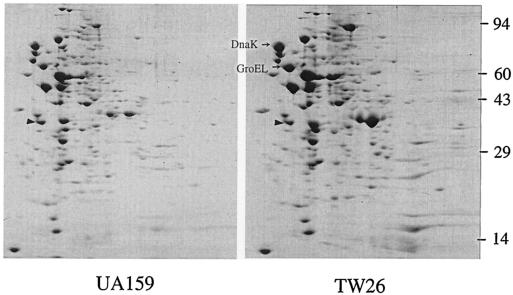
FIG. 7.
2D gel analysis. S. mutans UA159 and TW26 were grown in BHI medium to an OD600 of ∼0.5. Cell extracts of 200-μg total proteins were subjected to 2D electrophoresis using 2% ampholines at pH 4 to 8. Gels were stained with Coomassie blue. The triangles indicate the internal standard, tropomyosin, with pI 5.2 and a molecular weight of 32,700.

