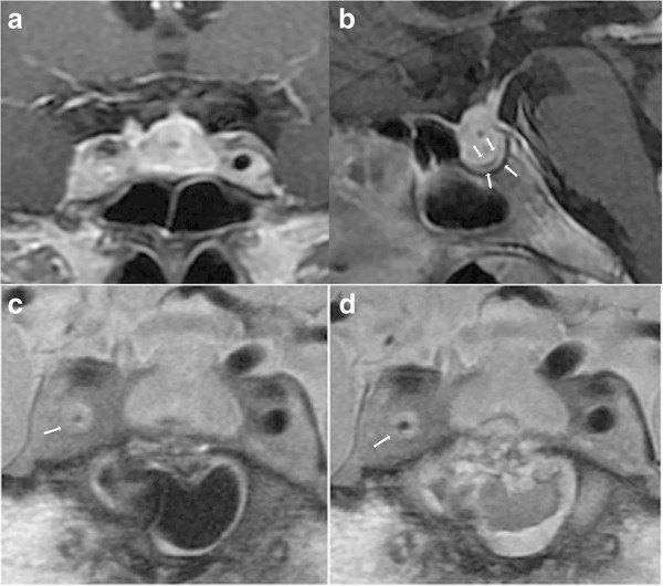
Figure 3.
Chronological MR images of Case 2. (a, b) Preoperative sagittal T1-weighted MR images with gadolinium revealing a sellar lesion extending to the suprasellar cistern (a), and markedly thickened dura mater (b). (c) Preoperative coronal plaque MR image revealing the thickened wall of the right internal carotid artery and the severely narrowed lumen. (d) Postoperative coronal plaque MR image showing improved right internal carotid artery stenosis.

