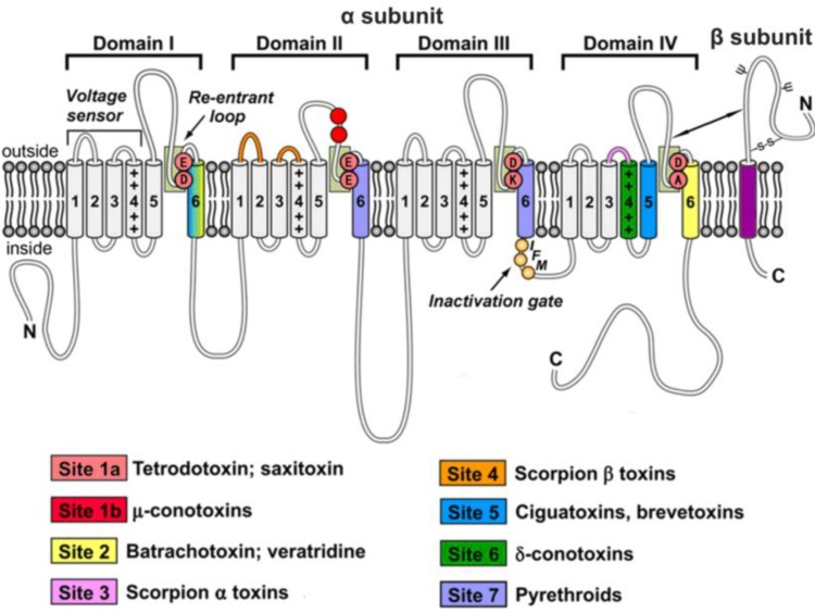
Figure 1.
Schematic diagram of molecular structure and pharmacology of VGSCs. VGSCs comprise a core protein α-subunit and one or more auxiliary β-subunits. The α-subunit consists of four homologous domains designated I–IV. Each domain is comprised of six transmembrane helical segments (S1–S6), which are represented by cylinders. The central pore is formed by the transmembrane segments S5 and S6, ion selectivity filter is formed by the segments SS1 and SS2 (re-entrant loops, the light green box), and the voltage-sensor is formed by the transmembrane segments S1 to S4. The positively charged S4 segment is principally responsible for sensing changes in membrane potential, modulating channels to open or close. The fast inactivation gate is formed by intracellular linker between transmembrane domains III and IV and contains an IFM (orange balls), which plugs the pore and prevents Na+ internal flow. Auxiliary β-subunits of VGSCs are illustrated in red cylinders. N-linked carbohydrate chains are presented by ψ. Different colored regions represent seven neurotoxin receptor sites. Figure adapted from King et al. (2008, 2012) and Catterall et al. (2007) [9,10,11].

