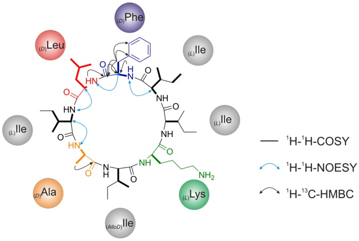
Figure 8.
NMR-spectroscopic structure assignment of champacyclin (1a) based on correlations between nuclei. Bold lines represent 1H-1H-COSY-, black arrows 1H-13C-HMBC- and blue arrows 1H-1H-NOESY-contacts. Proposed structure of (1a) is also based on data from chiral GC/PCI-EI-MS and HR-ESI-(+)-MSn analysis.

