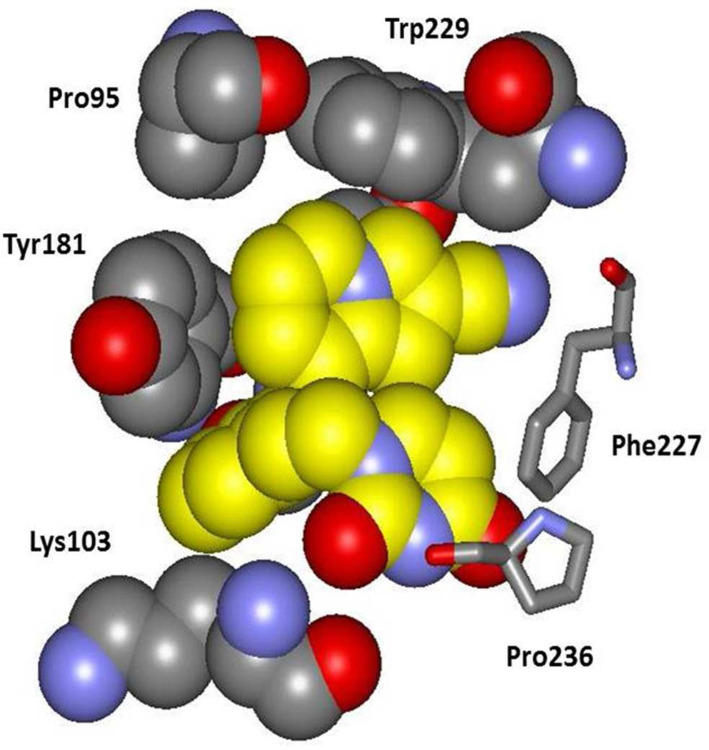
Figure 4.
A space-filling rendering made from the crystal structure of 10b bound to HV-RT. Carbon atoms of 10b are in yellow. Notable points are the hydrogen bond between a uracil oxygen atom and the backbone NH of Lys103, the aryl-aryl contacts with Tyr181 and Trp229, and the orientation of Lys103 including the contact of the Cβ atom with the inhibitor.

