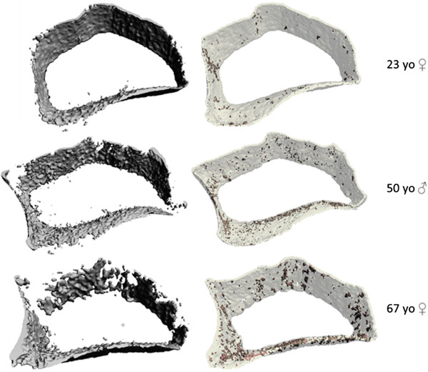
Figure 3.

Cortical compartment 3-dimentional reconstructed images. 3-D reconstructed images of segmented cortical compartments from the same HR-pQCT images of the ultra-ultra-distal radius region in three participants (top row - 23 y.o. female, middle row - 50 y.o. male, bottom row - 67 y.o. female) using the standard clinical evaluation protocol (left) compared to our semi-automated cortical segmentation protocol (right). The images on the right also show shaded areas of cortical porosity identified with the adapted direct transformation cortical evaluation script.
