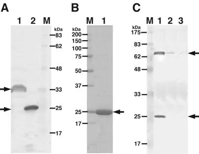
FIG. 1.
Electrophoretic profiles of Sphingomonas alginate lyases. (A) SDS-PAGE, followed by Western blotting with anti-His-tagged sequence antibodies. Lane 1, cell extract of E. coli transformed with pET21b-A1-II′(L); lane 2, cell extract of E. coli transformed with pET21b-A1-II′(S); lane M, molecular mass standards. (B) SDS-PAGE, followed by protein staining with Coomassie brilliant blue. Lane M, molecular mass standards (synthetic polypeptides); lane 1, purified A1-II′(S). (C) SDS-PAGE, followed by Western blotting with anti-A1-II′(S) antibodies. Lane M, molecular mass standards; lane 1, Sphingomonas sp. strain A1 cells grown on alginate; lane 2, cells grown on pectin; lane 3, cells grown on glucose. Arrows indicate alginate lyases.

