FIGURE 4.


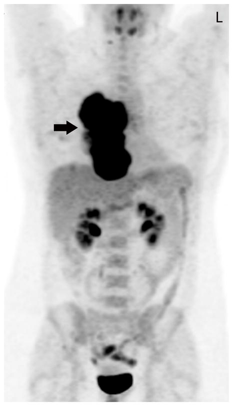
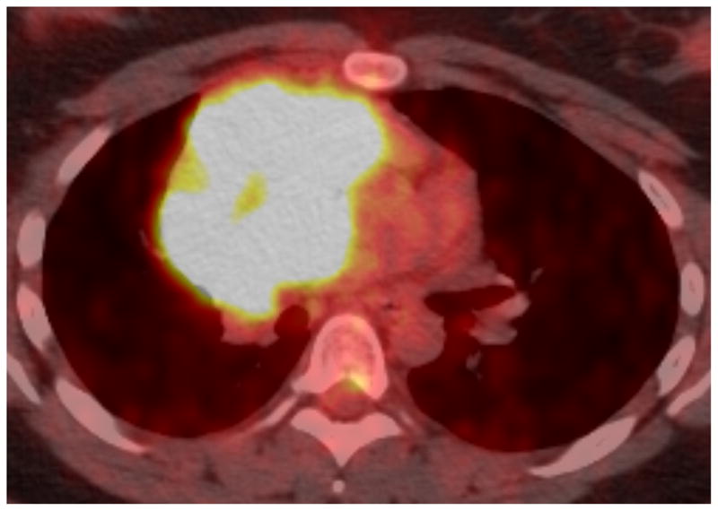
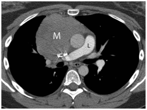
Twenty-nine-year-old woman complaining of chest pain over the last year. Contrast-enhanced chest CT image (A) at the level of the left pulmonary artery (L) demonstrates a large anterior mediastinal mass (M) deforming and surrounding the superior vena cava (*) for more than 180° of its circumference. Coronal maximum intensity projection FDG PET image (B) and fused axial PET-CT show the mass was intensely FDG avid, with a SUVmax of 15.5. Surgery confirmed a type B3 thymoma, stage IVa. The tumor directly involved the pericardium, superior vena cava, and right upper lobe, and there were small pleural metastases. CT, computed tomography. FDG, fluorodeoxyglucose. PET, positron emission tomography. SUV, standardized uptake value.

