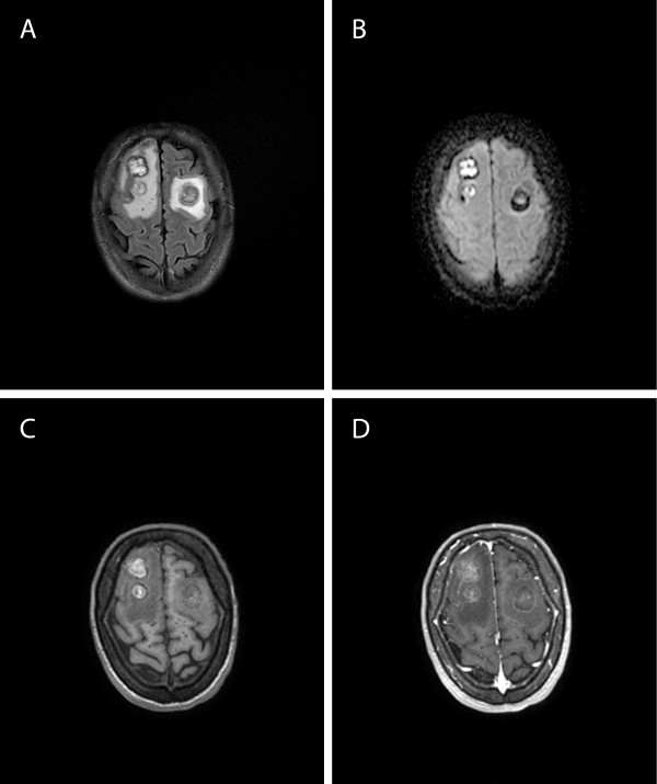
Figure 1.
Magnetic Resonance Images (MRI) of the brain at control after 26 days of anti-Toxoplasma treatment. A. On Axial FLAIR T2-weighted images the lesions are hyperintense, with hypointense halo and surrounding edema. B. Diffusion-weighted imaging of the lesions showed restricted diffusion. C. On unenhanced Axial T1-weighted images the lesions signal is heterogeneous. D. On enhanced Axial T1-weighted images faint lesions enhancement are depicted.

