Figure 1.

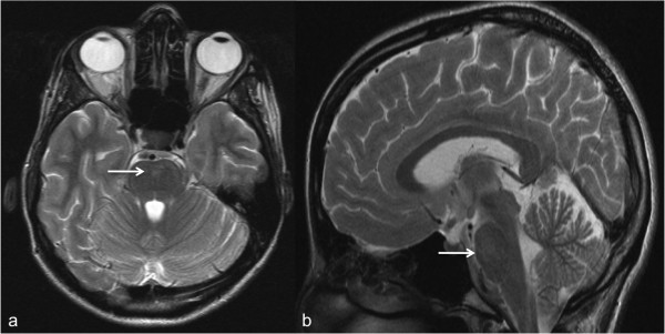
Brainstem abnormalities in a 28-year-old patient. (a) Axial and (b) sagittal T2w- TSE MR- images of the brain show hyperintense signal alterations in the pons (arrows). T2w, T2-weighted; TSE, turbo spin-echo; MR, magnetic resonance.

Brainstem abnormalities in a 28-year-old patient. (a) Axial and (b) sagittal T2w- TSE MR- images of the brain show hyperintense signal alterations in the pons (arrows). T2w, T2-weighted; TSE, turbo spin-echo; MR, magnetic resonance.