Figure 1.
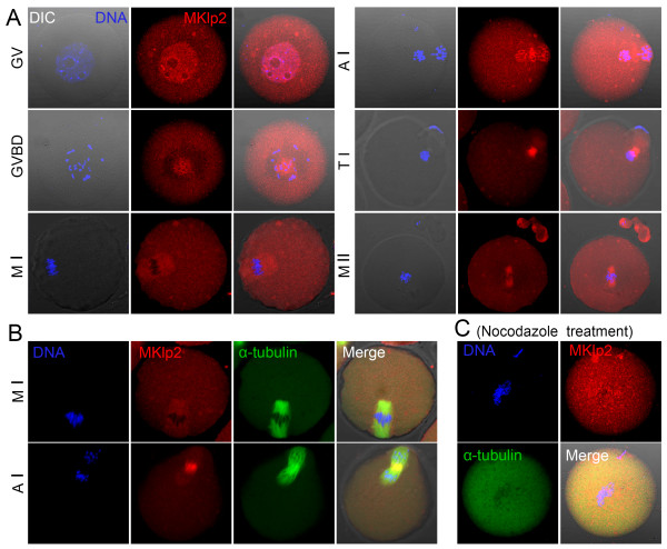
Expression and localization of MKlp2 in mouse oocytes. (A) Subcellular localization of MKlp2 during mouse oocyte meiotic maturation. MKlp2 antibody staining was employed to show the subcellular localization of MKlp2 in mouse oocytes. During mouse oocyte meiotic maturation, MKlp2 accumulated at the spindle and in the cytoplasm of oocytes. Red, MKlp2; blue, chromatin. (B) Co-localization of MKlp2 and α-tubulin. MKlp2 co-localized with spindle microtubules. Green, α-tubulin; red, MKlp2; blue, chromatin. (C) Localization of MKlp2 in mouse oocytes after treatment with nocodazole. Green, α-tubulin; red, MKlp2; blue, chromatin.

