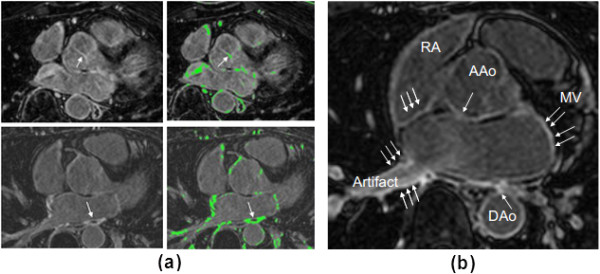
Figure 2.

Yale method’s threshold criterion. (a) Choice of threshold for pre-ablation (top) and post-ablation (bottom) images. Arrow points to fibrosis used for choosing threshold. Pre-ablation, this was a prominent section of the aortic valve. Post-ablation, the entire area of a prominent scar was selected. (b) The LA wall identification excludes regions where enhancement existed, but was attributed to artefacts or fibrosis of the mitral valve (MV), Aortic wall (AAo, DAo), or right atrial (RA) wall (arrows).
