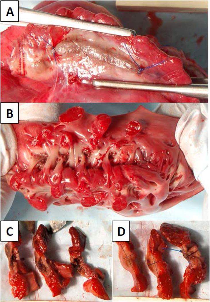
Figure 4.
Digital photographs demonstrating a representative ablation line from the (A) epicardial and (B) endocardial surfaces. After optimizing brightness and contrast, the digital photographs of the (C & D) cross-sections were marked for lesion depth and width. In total, 16 cross-sections were found to be non-trasmural (D, center cross-section).

