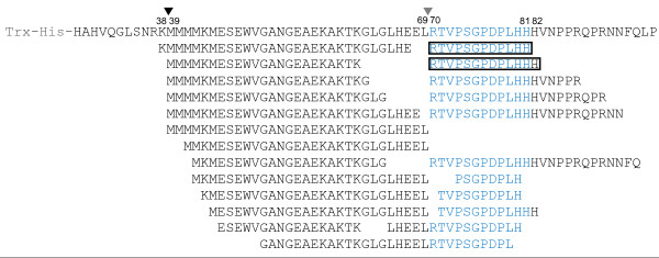
Figure 2.

Peptide fragments produced from ProCLV3 after incubations. Matching fragments detected are shown below the ProCLV3 sequence, as identified with Q-Tof MS/MS analyses. The core CLE motif (corresponding to the CLV3 peptide) of CLV3 is shown in blue. The CLV3p12 and CLV3p13 peptides are framed. Two internal cleavage sites, before Met39 and before Arg70, are indicated by arrowheads.
