
Figure 3.
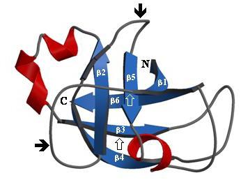
Modelled fold of a PNP-like molecule showing the six stranded double-psi β barrel structure. Fold recognition methods predict with certainty (Z score: >5) that AtPNP-A and the Xanthomonas axonopodis PNP-like molecule both adopt this fold. The N- and C-terminus of the protein are indicated, the α-helices are in red, the 6 β-strands are in blue and the two protruding psi loops are marked with a solid arrow (↑). The open arrows (↑) delineate the 33 amino acid long domain critical and sufficient for biological activity [5]. The N-terminal signal peptide that is not required for biological function outside the cell [5] was not included in the model. The model was generated using the software MOLSCRIPT [34].