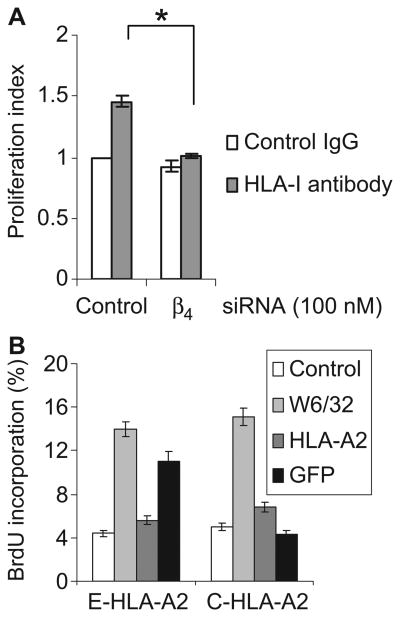
Fig. 5.
Integrin β4 is required for HLA-I–mediated cell proliferation. (A) Endothelial cells (EC1) were transfected with control or integrin β4–specific siRNA. Twenty-four hours later, cells were labeled with CFSE and stimulated with W6/32 for 72 hours. Cell proliferation was analyzed with ModFit LT software. The proliferation index represents the number of proliferating cells in test cultures as a ratio of the number of proliferating cells in the control cultures. Data are presented as mean proliferation index ± SEM from three independent experiments. *P < 0.05 by one-way ANOVA, Fisher's LSD. (B) Endothelial cells (EC2) were infected with adenoviruses expressing E-HLA-A2 or C-HLA-A2 and were stimulated with antibodies against GFP or HLA-A2 in the presence of BrdU overnight. Cells stimulated with mouse IgG or W6/32 served as negative and positive controls, respectively. Data are presented as the mean percentage of BrdU-positive cells ± SEM of three independent experiments. The flow cytometry plot of one representative experiment is presented in fig. S3.

