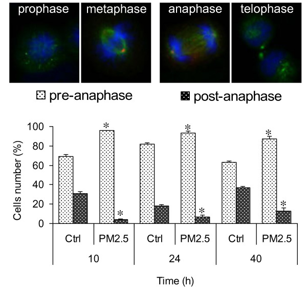
Figure 3.
Analysis of the mitotic phases. BEAS-2B cells were exposed for 10, 24 and 40 h to 7.5 μg/cm2 of winter PM2.5, stained for DNA (blue) and β-tubulin (green) and scored as pre-anaphasic and post-anaphasic cells. The results are representative of 3 independent experiments; in each experiment 200 cells were scored. *Statistically significant difference from untreated cells (control), P < 0.05.

