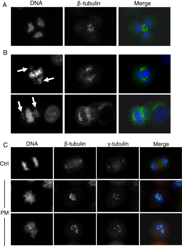
Figure 4.
Mitotic spindle analysis. BEAS-2B cells were exposed for 10 h to 7.5 μg/cm2 of winter PM2.5: β-tubulin (green) and DNA (blue) staining evidenced tripolar mitotic cell (A, telophase; 8% of mitotic cells in treated samples vs. 2% in controls; statistically significant difference, P < 0.05); and bipolar incomplete spindle with groups of lagging chromosomes (B, arrows; 10% of mitotic cells in treated samples vs. 1% in controls; statistically significant difference, P < 0.05); γ-tubulin staining (red) showed centrosomes amplification (C, 6.7% of mitotic cells in treated samples vs. 2.7% in controls; statistically significant difference, P < 0.05). The results are representative of 3 independent experiments; in each experiment 300 cells were scored.

