Figure 3.
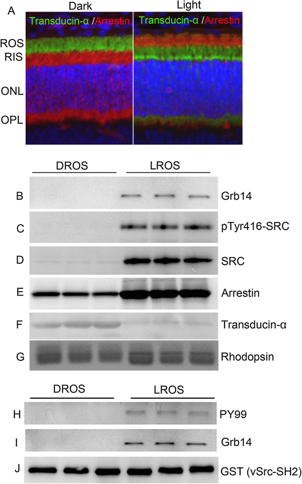
Association of Grb14 and Src to light-adapted rod outer segment membranes. Immunofluorescence analysis with anti-transducin (Tα) and anti-arrestin antibody was performed with dark- and light-adapted (300 lux for 30 min) rat retinal sections (A). The images are of the same section viewed with a filter to detect transducin α (green), arrestin (red), and DAPI stained nuclei (blue). ROS, rod outer segment; RIS, rod inner segment; ONL, outer nuclear layer; OPL, outer plexiform layer. ROS membranes were prepared from dark- and light-adapted rats on a discontinuous sucrose density gradient centrifugation. DROS and LROS proteins were subjected to immunoblot analysis with anti-Grb14 (B), anti-pTyr416-Src (C), anti-Src (D), anti-arrestin (E), anti-transducin-α (F), and anti-rhodopsin (G) antibodies. Solubilized DROS and LROS proteins were incubated with the GST-vSrc-SH2 fusion protein for 2 h followed by GST pull-down assay. The bound proteins were subjected to immunoblot analysis with anti-PY99 (H), anti-Grb14 (I), and anti-GST (J) antibodies.

