Figure 2.

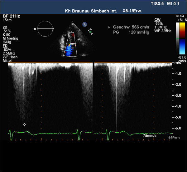
Two-dimensional color-flow Doppler echocardiogram shows blood flow during systole from the left ventricle to the right atrium through the Gerbode defect (arrows) in apical 4-chamber view.

Two-dimensional color-flow Doppler echocardiogram shows blood flow during systole from the left ventricle to the right atrium through the Gerbode defect (arrows) in apical 4-chamber view.