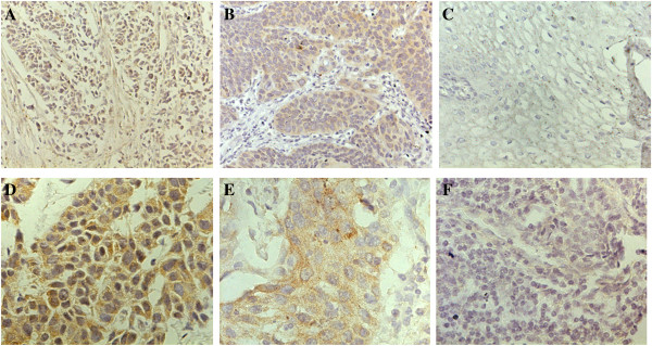
Figure 1.

Immunohistochemical expression of CypA and MMP9 in esophageal squamous cell carcinoma. A, D Typical immunohistological features with high levels of CypA expression in esophageal squamous cell carcinoma (ESCC). The CypA staining shown nuclear and cytoplasmic localization; B, E Typical immunohistological features with high levels of MMP9 in ESCC. The MMP9 staining was present in the cytoplasm of tumor cells; C, F Negative staining in ESCC. Magnifications: A-C × 200, D-F × 400.
