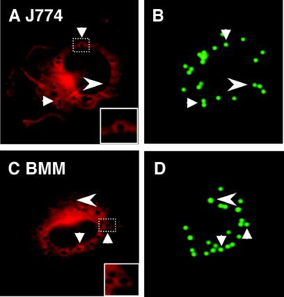
FIG. 2.
Recruitment of iNOS to model, LB phagosomes: J774 macrophages (A and B) and BMM from C57BL/6 mice (C and D) stimulated with IFN-γ and LPS. After phagocytosis of LB, cells were stained with anti-iNOS antibodies followed by a secondary antibody conjugated to Alexa 568. Arrows indicate examples of iNOS colocalization with LB phagosomes. Arrowheads indicate LB phagosomes that did not colocalize with iNOS. Insets (A and D) are enlarged images corresponding to the dashed boxes, as indicated.

