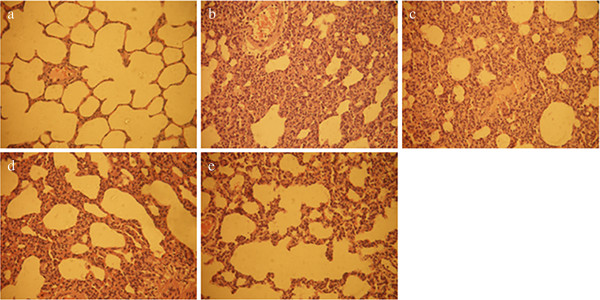
Figure 1.

Lung pathologies. (a) Control group, (b) L group, (c) TP1 group, (d) TP2 group, and (e) TP3 group with hematoxylin-eosin (H&E) staining (×400). Injection of LPS caused infiltration of inflammatory cells into the lung interstitial and alveolar spaces, alveolar wall thickening, and intra-alveolar exudation. In TP1, TP2, and TP3 groups, triptolide attenuated these histological changes. LPS, lipopolysaccharide.
