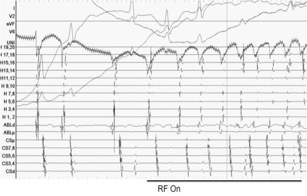
Figure 4.

Intracardiac signals. Displayed are surface ECG, Unipolar EGM, Halo-catheter EGM (H1-H20), ablation catheter and coronary sinus EGM (CS 1-10). The Halo catheter was positioned circumferentially (roof (H15-20)>lateral wall (H5-14)>floor (H1-4)) around the right atrium with some poles straddling the crista terminalis (double potentials). The recording demonstrates a QS early signal on unipolar mapping together with tachycardia acceleration at the onset of RF ablation.
