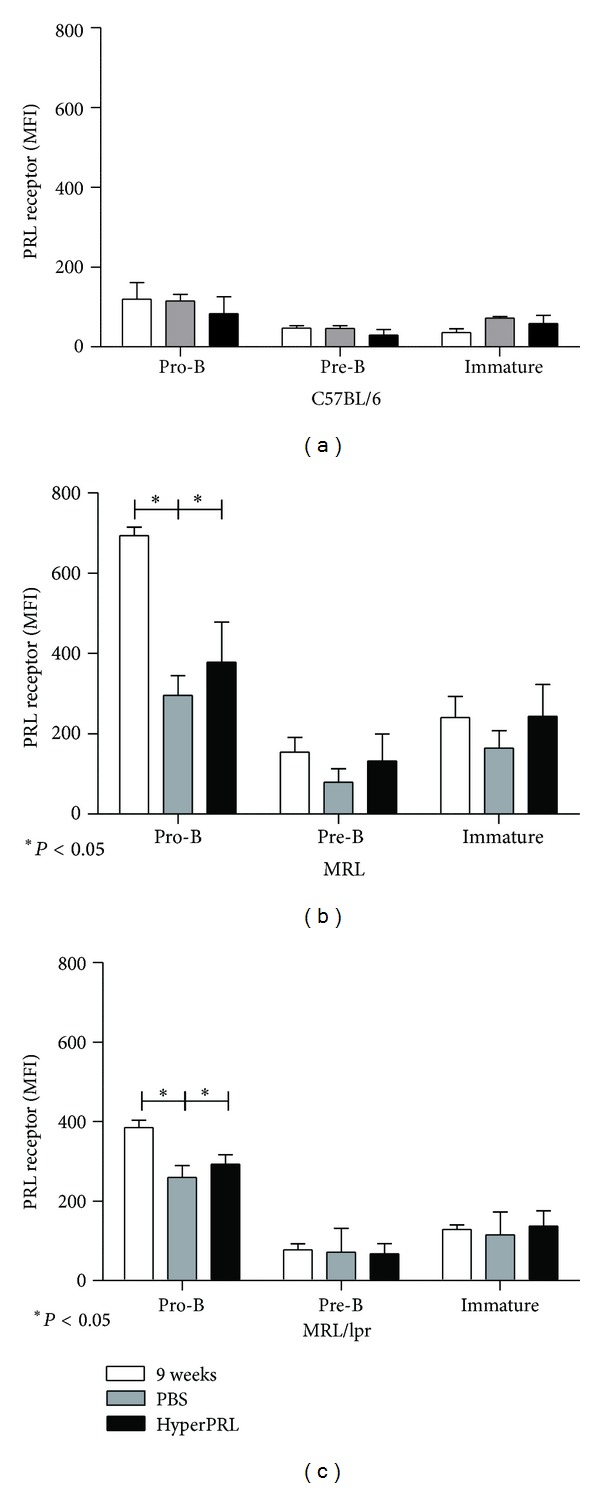
Figure 3.

Prolactin receptor expression after the induction of hyperprolactinemia. The levels of PRL receptor protein (MFI) in the B cells from BM (pro-B, preB, and immature) were measured using flow cytometry. At the end of the treatment, the BM cells were labelled with anti-B220, anti-CD43, anti-CD23, anti-IgM, and goat anti-PRL receptor antibodies. (a) C57BL/6 mice; (b) MRL mice; (c) MRL/lpr mice. The asterisks denote statistical significance between populations with the P value shown. The MFI values expressed in the graphs correspond to the MFI values minus the isotype control.
