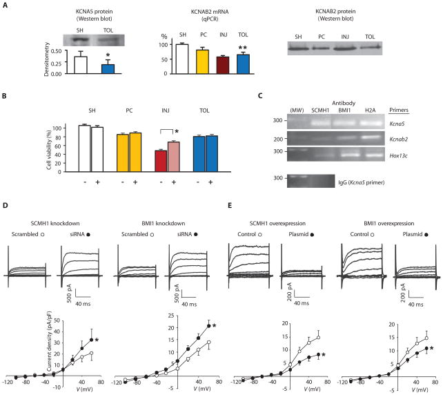
Fig. 4.
Implication of potassium channel suppression in ischemic tolerance and inhibition of K+ current by PcG proteins. (A) Analyses of potassium channel abundance in brain. Left, KCNA5; middle and right, KCNAB2. SH, PC, INJ, and TOL: as noted in Fig. 1. (B) Effects of knocking down potassium channels on tolerance. NS20Y cells were transfected with scrambled RNA (−) or a mixture of siRNA oligos (+) against Kcna5 and Kcnab2, followed by differentiation and OGD treatments as described in Figs. 2B and 3, A and B. Data are presented as mean ± SE of at least three independent cultures. (C) Results of ChIP assays of NS20Y cells, with antibodies noted in the figure and PCR primers, also noted in the figure, for the promoter region of Kcna5, Kcnab2, or Hox13c. Predicted sizes for PCR fragments are 270, 184, and 271 base pairs for Kcna5, Kcnab2, and Hox13c, respectively. Antibody against H2A and Hox13c primers were included as positive controls and immunoglobulin G (IgG) as a negative control. The analyses were repeated twice on two independent cultures with similar results. (D and E) Effects of knocking down (D) or overexpressing (E) SCMH1 or BMI1 on potassium currents. NS20Y cells were transfected with siRNA oligos or plasmids as noted in the figure and differentiated (n = 6 to 8 each condition). *P < 0.05; **P < 0.005.

