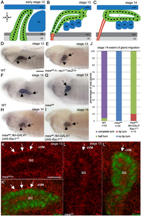
Fig. 1.
αPS1βPS acts upstream of Rac1 GTPase to control salivary gland migration. In wild-type embryos at early stage 12 (A), the distal tip of the salivary gland contacts the surrounding cVM and SM and turns posteriorly although the proximal cells have not migrated dorsally (A). During stage 13, distal cells of the wild-type gland continue to turn posteriorly as they extend basal membrane protrusions that contact surrounding tissues while the proximal cells migrate dorsally (B). By stage 14 the wild-type gland has turned completely (C). In stage 13 wild-type embryos (D), the distal half of the salivary gland turns posteriorly (D, arrow) while the proximal cells migrate dorsally D, arrowhead, whereas at stage 14 (F), the distal (F, arrow) and proximal (F, arrowhead) gland cells have completed the posterior turn. In stage 13 embryos trans-heterozygous for mewM6 and rac1J11rac2Δ (E) and stage 14 mewM6 homozygous embryos (G), the distal salivary gland cells do not turn posteriorly (E and G, arrows) although the proximal cells migrated dorsally (E and G, arrowheads). Expression of activated Rac1V12 in all salivary gland cells of mewM6 mutant embryos with fkh-GAL4 (H and I) results in posterior turning of the distal cells (H and I, arrows) although the gland does not continue migrating and the proximal cells do not turn (E and F, arrowheads). Graph depicting extent of salivary gland migration in stage 14 wild-type embryos, mewM6 mutant embryos and mewM6 mutant embryos expressing Rac1V12 in the gland (J). In mewM6 heterozygous embryos (K), endogenous Rac1 localizes to sites of contact between the basal membrane of salivary gland cells and overlying cVM (K and K′, arrows), whereas in homozygous siblings (L), Rac1 does not localize to the contact site (L and L′, arrows). Embryos in D-I were stained for dCREB with embryo in E being stained for β-galactosidase (β-gal; not shown) to distinguish rac1J11rac2Δ heterozygous from homozygous embryos. Embryos in K and L were stained for dCREB (green) and Rac1 (red). Scale bar in A represents 20 μm and the scale bar in H represents 10 μm. cVM: circular visceral mesoderm; SM: somatic mesoderm; NB: neuroblasts; VE: ventral ectoderm; FB: fat body; D: dorsal; V: ventral; A: anterior; P: posterior.

