Fig. 7.
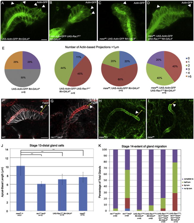
Rac1 is required for basal membrane protrusions in the distal salivary gland. Distal salivary gland cells expressing Actin-GFP in wild-type embryos (A) and mewM6 homozygous embryos expressing Rac1V12 in the gland (D) extend basal membrane protrusions (A and D, arrowheads). Distal salivary glands expressing Rac1N17 and Actin-GFP (B) or glands of mewM6 homozygous embryos expressing Actin-GFP (C) do not extend basal membrane protrusions (B and C, arrowheads). Pie-charts show percentage of glands with indicated number of basal membrane protrusions per gland in wild-type glands expressing actin-GFP in the gland, glands co-expressing Rac1N17and actin-GFP, mewM6 mutant glands expressing actin-GFP in the gland and mewM6 mutant glands co-expressing Rac1V12 and actin-GFP (E). In wild-type embryos (F) and mewM6 heterozygous embryos (H), distal gland cells are elongated (F and H, arrowheads) whereas in rac1J11rac2Δ (G) and mewM6 (I) mutant embryos, distal gland cells are not elongated (G and I, arrowheads). Graph shows apical-basal length of distal gland cells in mewM6 heterozygous embryos, rac1J11rac2Δ and mewM6 mutant embryos and embryos expressing Rac1N17 in the gland (J). Graph shows extent of salivary gland migration at stage 14 in rac1J11rac2Δ heterozygous and homozygous embryos and wild-type and rac1J11rac2Δ embryos expressing Rac1F37A or Rac1Y40C (K). Panels A–D are live-images of embryos expressing Actin-GFP specifically in the salivary gland with fkh-GAL4. Embryos in panels F and G were stained for E-cad (red), FasIII (white) and β-gal (not shown). Embryos in H and I were stained for F-actin (green). All embryos shown are at stage 13. Scale bar represents 10 μm.

