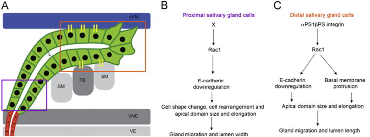
Fig. 8.
Model for αPS1βPS and Rac1 function in migration of distal and proximal salivary glands. Schematic diagram of a migrating salivary gland at stage 13 (A) showing the proximal gland cells (purple box) and distal gland cells (orange box) that interact with the overlying cVM (blue) and underlying FB/SM (gray) through integrin adhesion receptors (yellow sticks). In the proximal gland cells (B), an unknown factor X activates Rac1 to downregulate E-cadherin and control cell shape change, rearrangement and apical domain size and elongation which are important for dorsal migration of the cells and lumen (L) width. In the distal gland cells (C), αPS1βPS activates Rac1 to downregulate E-cadherin and promote basal membrane protrusion which allows correct apical domain size and elongation and are important for gland migration and lumen length.

