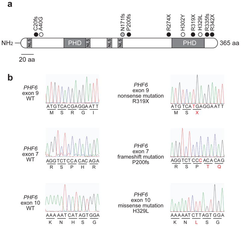
Figure 1. PHF6 mutations in AML.
(A) Schematic representation of the PHF6 protein depicting the location of 4 nuclear localization signals and 2 imperfect PHD zinc finger domains. Overview of PHF6 mutations identified in primary AML samples. Filled circles represent nonsense and frameshift mutations, whereas missense mutations are depicted as open circles. The circle filled in gray indicates a frameshift PHF6 mutation identified in a female AML sample. (B) Representative DNA sequencing chromatograms of AML DNA samples showing mutations in exons 7, 9 and 10 of PHF6.

