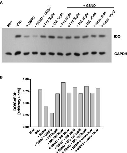
FIG. 6.
Proteasome inhibitors abolish NO-dependent IDO degradation. A549 cells were stimulated with 50 U of IFN-γ per ml for 9 h. Thereafter, the cells were washed, and the proteasome inhibitors MG-132, proteasome inhibitor I (PSI), and clasto-lactacystin β-lactone (clasto) were added at the indicated final concentrations to fresh culture medium. The addition of dimethyl sulfoxide to a final concentration of 0.5% served as a solvent control (GSNO plus DMSO, lane 4), and unstimulated cells as a negative control (Med, lane 1). One hour later, the NO donor GSNO was added to a final concentration of 750 μM, and the cells were further incubated for 16 h. IDO and GAPDH protein levels were determined by Western blotting (A). The signal intensity of the IDO bands was measured by densitometry and normalized to the intensity of the corresponding GAPDH signal. The results shown are representative of one of three independent experiments (B).

