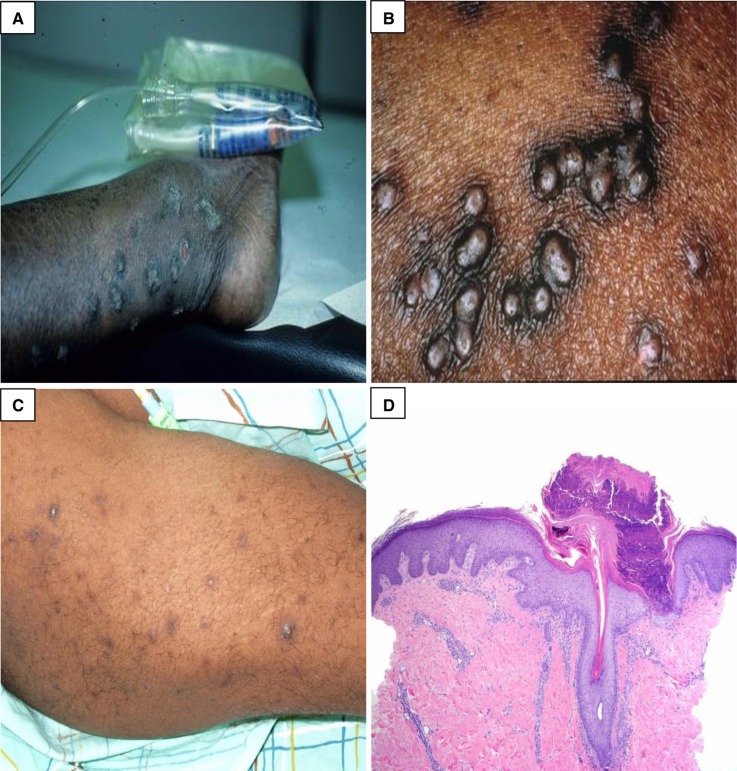
Figure 2.
Acquired perforating dermatosis. (A and B) Multiple clustered, hyperpigmented, dome-shaped nodules and coalescing plaques with a central keratotic plug. (C) Hyperpigmented macules and patches from lesions that have resolved. (D) There is a cup-shaped epidermal depression filled with parakeratosis and neutrophilic debris. At the base of the depression, the epidermis is thinned, and degenerated collagen fibers are noted protruding through this attenuated epidermis. (Courtesy of Timothy G. Berger, Anna K. Haemel, and Thaddeus W. Mully.)

