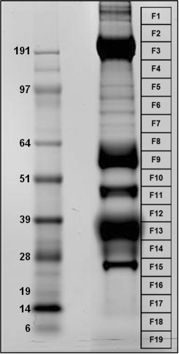
Figure 1.

Salivary gland proteins from the mosquito Psorophora albipes. The left gel lane shows the protein standards with their molecular weights (kDa). The right gel lane shows the P. albipes salivary proteins (Coomassie stained). The grid at the right (F1–F19) shows the area of the gel slices submitted for tryptic digest and tandem mass spectrometry identification.
