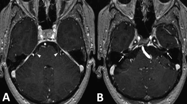
Figure 1.

Axial MRI images of the brain. Post gadolinium enhanced T1 weighted sequence on initial exam shows (A) bilateral focal enhancement of the proximal portion of both trijeminal nerves (arrowheads), (B) bilateral enhancement of the vestibulocochlear and facial nerves, more pronounced on the right side (arrow) and bilateral thickening of the Gasser’s ganglia walls (*).
