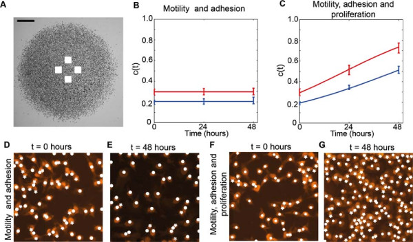
Figure 7.

Cell density measurements where cell proliferation is not suppressed allows us to estimate λ. The approximate location of the subregions used to measure the cell density are shown in (A), where the scale bar corresponds to 1.5 mm. Images in (D–E) show two subregions of dimensions 230 μm× 230 μm for experiments at t=0 hours and t=48 hours, where 30,000 cells, pretreated with Mitomycin–C, were initially placed inside the barrier. Equivalent images without Mitomycin–C pretreatment are shown in (F–G). The Propidium Iodide staining is highlighted in orange. For each subregion, the number of cells was counted; white circles correspond to the cells automatically detected by the image analysis software and white stars indicate cells that were manually counted. The corresponding time evolution of the mean scaled density, c(t), is shown in (B) and (C), where the error bars indicating one standard deviation from the mean. Blue and red data points correspond to the experiments initialised with 20,000 and 30,000 cells, respectively. Our analysis shows that the proliferation rate (λ) and the doubling time (td=loge2/λ) for the experiments initialised with 20,000 cells is λ=0.0305hour−1 and td=22.7 hours, and for experiments initialised with 30,000 cells is λ=0.0398hour−1 and td=17.42 hours. The red and blue curves in (B) and (C) show the corresponding solution of the logistic equation, given by Equation 2, respectively.
