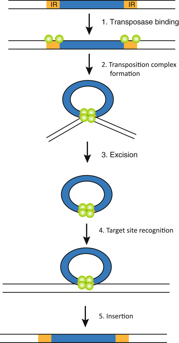
Figure 1.
Model of cut-and-paste transposition. Transposase proteins (green spheres) recognize the terminal inverted repeats (IRs, orange boxes) and form a circular pre-excision synaptic complex from which the transposon is excised. Formation of the synaptic complex, allowing close association of the two transposon ends, involves the formation of transposase tetramers [74]. Subsequently, the transposition complex recognises a target site and the transposase proteins mediate integration of the transposon. The figure shows the proposed mechanisms for Tc1/mariner-type (e.g. Sleeping Beauty) DNA transposition. Yellow boxes marked with IR represent the terminal inverted repeats.

