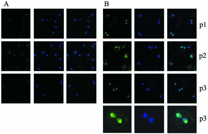
FIG. 3.
Immunofluorescence reactivity of antiserum against BbAMA-1 incubated with acetone-fixed B. bovis-infected bovine erythrocytes. (A) Incubation with preimmune sera. (B) Incubation with immune sera against peptide 1 (p1), peptide 2 (p2), and peptide 3 (p3), as indicated on the right. Panels in the left column show anti-AMA-1 staining; panels in the middle column show DAPI staining; and panels in the right column are overlay images. The images in the bottom row of panel B are enlargements of duplicated B. bovis merozoites reacting with anti-peptide 3.

