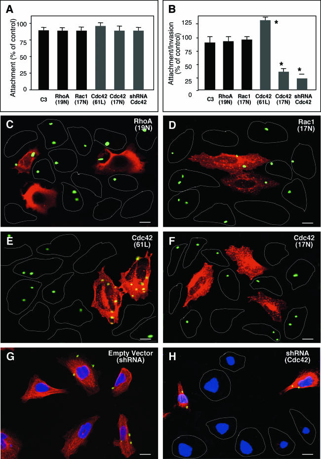
FIG. 3.
C. parvum invasion of biliary epithelial cells requires the activation of host cell Cdc42, but not RhoA and Rac1. Cells were transfected with either a consitutively active mutant of Ccd42 or dominant negative mutants of Cdc42, RhoA, or Rac1 and then exposed to C. parvum followed by immunofluorescent microscopy. Some cells were transfected with a vector encoding an shRNA toward Cdc42 before exposure to C. parvum. (A) Attachment assay in prefixed cells after a 2-h exposure to C. parvum sporozoites shows no significant difference of C. parvum attachment in all the treated cells. (B) Attachment-invasion assay in nonfixed cells after a 2-h exposure to C. parvum sporozoites. A significant increase of infection rate was found in cells transfected with Cdc42 (Q61L) and a significant decrease of infection rate was detected in cells transfected withCdc42 (17N) or Cdc42 siRNA, but not RhoA (19N) and Rac1(17N) or cells treated with exoenzyme C3. (C to H) Representative confocal images of cells of various treatment exposed to C. parvum for 2 h. Transfected cells were identified by immunostaining using an antibody to the C-Myc epitope tag. No significant different of C. parvum infection was found between nontransfected cells as outlined (C and D) and cells transfected with RhoA (19N) (stained in red in C) or Rac1 (19N) (in red in D). More C. parvum parasites were detected in cells transfected with Cdc42 (61L) (in red in E) and much fewer C. parvum parasites were found in cells transfected with Cdc42 (17N) (in red in F) compared with nontransfected cells (as outlined in E and F). Whereas cells transfected with the empty shRNA vector displayed a normal Cdc42 cellular expression and a similar infection pattern as nontransfected cells (G), cells transfected with shRNA to Cdc42 showed no obvious expression of Cdc42 (with nucleus staining with DAPI but absence of Cdc42 expression in H) and a marked decrease of C. parvum infection compared with nontransfected cells (shown expression of Cdc42 in H). *, P < 0.05 (compared with no-inhibitor treated or nontransfected controls); error bars, standard errors of the means; scale bars = 5 μm.

