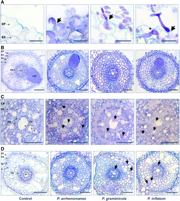
Figure 2.
Histological study of the Pythium infection process in rice roots. A, Direct penetration of the rice rhizodermis by P. arrhenomanes, P. graminicola and P. inflatum. Arrows indicate sites of hyhal penetration. Bulbous-like Pythium hyphae grew intracellularly and became severely constricted right before cell wall passing (*). B, Colonization of the inner rice root tissues by P. arrhenomanes, P. graminicola and P. inflatum in the most heavily infected (upper) parts of the primary root at 27 hpi. C, Colonization of the stele by P. arrhenomanes, P. graminicola and P. inflatum in the most heavily infected parts of the primary root at 27 hpi. Arrows indicate hyphae in the phloem and xylem. D, Colonization of the inner root tissues 2 days upon inoculation with P. arrhenomanes, P. graminicola and P. inflatum in the middle part of the primary root. P. graminicola and P. inflatum hyphae were less abundant in the cortex and vascular tissues (arrows). Abbreviations in the control pictures designate the epidermis (EP), exodermis (EX), sclerenchyma (SC), cortex (C), endodermis (EN), pericycle (PE), stele (ST), phloem (PH), xylem (XY) and lateral root primordium (LRP). Cross-sections (5 μm) from Pythium-infected root tissues were stained with trypan blue. Scale bars in A = 20 μm, in B = 100 μm, in C = 50 μm and in D = 100 μm.

