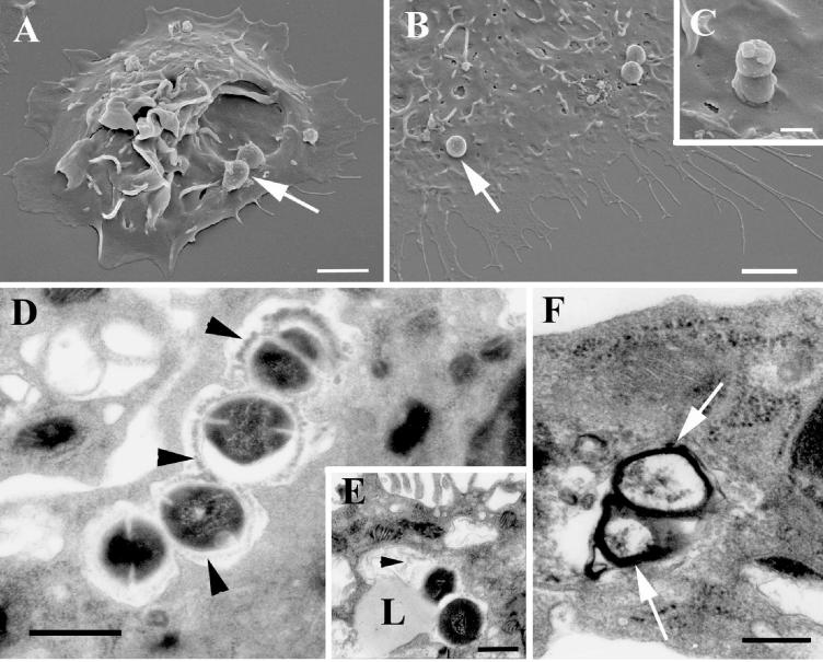
FIG. 3.
Electron microscopy examination of S. pyogenes-infected peritoneal macrophages. (A to C) Scanning electron microscopy revealed streptococci (arrows) attached to macrophages (A and B) and microorganisms in the process of being internalized (C). Ultrathin sections of infected macrophages show degradation of intracellular streptococci. (D) The capsular structure (arrowheads) is being detached from the surface of the bacterium and undergoing degradation. (E) Fusion of a lysosome (L) with the phagosome. Again, degraded capsular material (arrowhead) can be identified in the phagolysosome. (F) This process leads to a complete degradation of the microorganism. Only the debris of the cell wall (arrows) can be found inside the phagolysosome. Bars, 2 (A and B), 0.5 (C), and 1 (D to F) μm.

