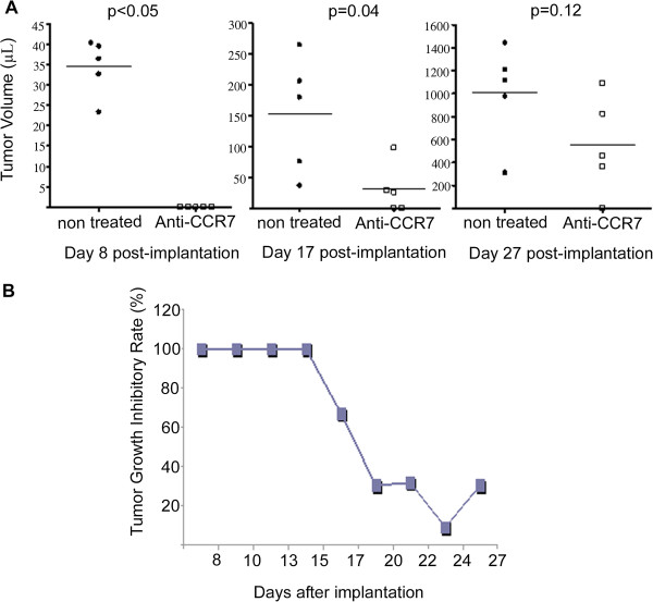
Figure 2.
Anti-CCR7 mAb reduces tumor growth of MCL cells. Granta-519 cells were implanted subcutaneously in the right flank of NOD/SCID mice (n = 10). Treatment started at day 2 post-inoculation and continued at days 6 and 10. Anti-CCR7 mAb were administered intraperitoneally. Once subcutaneous tumors were palpable (≥4 mm) the diameters of the tumor mass was measured every 3 days. (A) Effect of anti-CCR7 mAb treatment on tumor volume in Granta-519 xenograft-bearing mice. An altered growth pattern in the treated and control groups are shown in the graph. The time points correspond to day 8 (first palpable tumors), day 17 and day 27. Each black dot (●, control group) or white square (□, treated group) represents one individual mouse. Horizontal bars represent mean tumors volume of each group, which consisted of 5 mice. Tumor volumes were calculated according to the V = D * d2/2 formula, as stated in Design and methods. P-values refer to the tumor volume differences from mice treated with PBS or anti-CCR7 mAb at different time points. (B) Anti-CCR7 mAb delays the development of subcutaneous tumors. The maximum growth inhibitory rate (IR) was reached within the first two weeks post anti-CCR7 mAb administration. The inhibitory rate is shown and is given by IR(%) = 100 ‒ [(V1/V2)] * 100 where V1 is the mean tumors volume in the mAb treated groups and V2 is the mean tumors volume in the control group.

