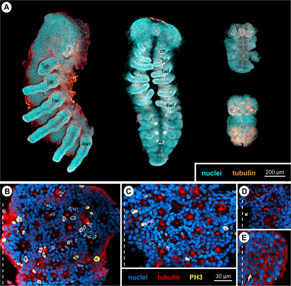
Figure 16.
Comparison of germ band size, cell number and cell sizes between callipallenid pycnogonids and spiders. Tubulin-labelled embryos with nuclear counterstain, in part with additional PH3 labelling. (A) Ventral view of germ bands of Cupiennius salei (left, only left half of prosomal region shown) and Parasteatoda tepidariorum (middle) compared to Callipallene sp. (right-top) and Pseudopallene sp. (right-bottom), flat preparations, Imaris volumes. (B–E) Apical horizontal sections (2D projections of curved composite optical sections). Dashed line marks the VMR. (B)C. salei, hemi-neuromere of walking leg segment 2. Note the apical mitoses scattered between the CISs. (C)P. tepidariorum, hemi-neuromere of walking leg segment 2. Note some scattered apical mitoses and the distinct tubulin labelling of CISs. (D)Callipallene sp., hemi-neuromere of walking leg segment 1. The apical area of the hemi-neuromere represents not even a quarter of the area in the corresponding hemi-neuromeres of the two spider species. Neuroectodermal cell size appears similar to spiders. Cross marks damaged region. (E)Pseudopallene sp., hemi-neuromere of walking leg segment 1. The apical area of the hemi-neuromere is significantly smaller and the neuroectodermal cells are slightly larger than in the two spider species.

