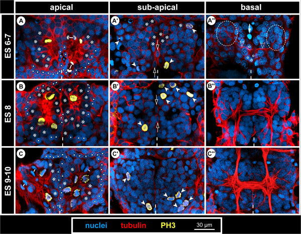
Figure 8.
Structure and mitoses in advanced walking leg neuromere 1 of Pseudopallene sp. (late ES 6 – ES 10) – part I. Optical sections of tubulin- and PH3-labelled embryos with nuclear counterstain. Dashed vertical line at the bottom of each image indicates the VMR. Small white lines connect newly forming nuclei during telophase. (A–C) Apical horizontal sections. Large apical NSCs line the paired invagination (asterisks), some of them being in division. Prior to hatching (C) compression of the apical hemi-ganglion portion by the outgrowing limb anlagen leads to a squeezed appearance of the invaginations and cells. Smaller prospective epidermal cells (white spots) line the rim of the invaginations. (A’–C’) Sub-apical horizontal sections. Divisions of sub-apical INPs (arrowheads) are observable throughout this phase of embryonic neurogenesis, being predominantly located in the anterior and posterior regions of the hemi-ganglion anlagen. Open arrows mark unpaired median cells of the VMR (putative glial cells). (A”–C”) Basal horizontal sections. Axonogenesis starts in late ES 6 (A”), the connectives being pioneered (not shown) and a commissural pathway (open arrowheads) being established by neurons located in the antero-lateral basal region of the hemi-ganglion anlage (stippled ovals). Medially, the growth cones have not yet crossed to the contra-lateral side and are in close contact to some of the basal cells of the VMR (open spots). Arrow marks a pycnotic body. During subsequent embryonic development (B”,C”) the slender transverse pathway differentiates into a compact undivided segmental commissure.

