Figure 1.
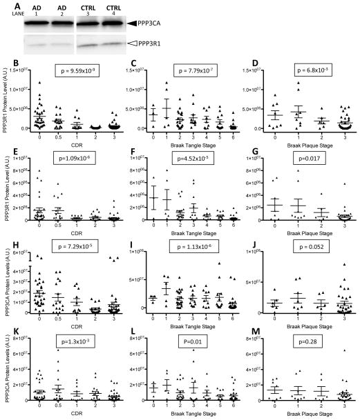
Calcineurin protein levels are altered in AD brains. Human brain samples from AD patients (N=73) and non-demented controls (N=39) were extracted in non-ionic detergent. A. Total cell lysate extracted from brains (100 ug) were analyzed by SDS-PAGE and immunoblots were probed with antibodies to the regulatory (PPP3R1) and catalytic (PPP3CA) subunits of calcineurin. B–M. Quantification of immunoblots. B–D and H–J. To correct for total protein, PPP3R1 and PPP3CA protein levels are expressed relative to β-tubulin protein levels. E–G and K–M. To correct for neuron number, PPP3R1 and PPP3CA protein levels are expressed relative to Tuj1 protein levels. B–G. Calcineurin regulatory subunit (PPP3R1) protein levels are associated with clinical and pathologic measures of AD. B,E. PPP3R1 protein levels are plotted with CDR. C,F. PPP3R1 protein levels plotted with Braak tangle staging. D,G. PPP3R1 protein levels are plotted with Braak plaque stage. H–M. Protein levels of the catalytic subunit of calcineurin (PPP3CA) are associated with clinical and pathologic measures of AD. H,K. PPP3CA protein levels are plotted with CDR. I,L. PPP3CA protein levels are plotted with Braak tangle staging. J,M. PPP3CA protein levels are plotted with Braak plaque staging.

