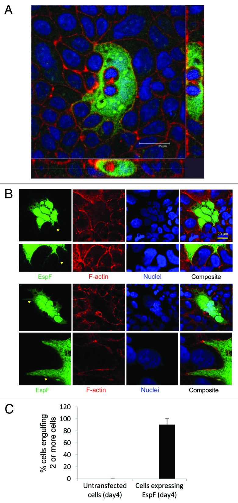
Figure 3. EspF expression induces cell internalization in an epithelial monolayer. (A) TC-7 cells expressing EspF(L16E)-GFP were visualized by confocal microscopy and sectioned along the x-z axis. (B) Cytoplasmic extensions (yellow arrow) in EspF-expressing cells (day 4 post transfection) were consistently found surrounding non-transfected cell types. (C) Quantification of cell engulfment in which only cells engulfing 2 or more cells were included in the analysis. Data shows mean ± SD, n = 3.
