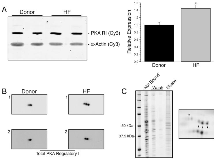
Fig. 4.
Expression of type I PKA Regulatory subunits. In failing myocardium, the relative expression of RIα significantly increased by 45% over donor samples (A, *P < 0.05 vs. donor). To scout for possible post-translational modifications of RIα, 2D Western blotting was done on two different donor and two different heart failure samples and showed up to three isoelectric variants of RIα (B). The presence of three isoelectric variants was additionally confirmed by capture of cAMP-binding proteins from a homogenate prepared from the HF1 sample by 8-AEA-cAMP-agarose beads (C, left). The fraction of the homogenate not bound to beads, the subsequent washes and the eluted fraction from the beads were analyzed by 1D SDS–PAGE and silver staining. Subsequently, the eluted fraction was analyzed by 2D SDS–PAGE and silver staining (C, right) which recapitulated the observed RIα pattern (denoted by arrowheads) as well capturing RIIα (denoted by *).

