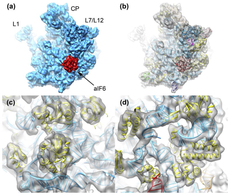
Fig. 2.

The 3D reconstruction of the M. thermautotrophicus 50S ribosomal subunit. (a) The cryo-EM structure of the M. thermautotrophicus 50S subunit (cyan) reveals density for aIF6 (red) bound in the sarcin–ricin loop region (L1: L1 stalk; L7/L12: L7/L12 stalk; CP: central protuberance). (b) The reconstruction of the M. thermautotrophicus 50S subunit (transparent gray) fitted with molecular structures and models for aIF6 and the ribosomal subunit. See the text for details. (c and d) The 6.6-Å cryo-EM density shows features typical for subnanometer-resolution cryo-EM reconstructions, such as rRNA major and minor grooves and tubular density for protein α-helices.
