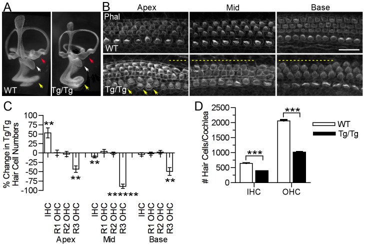
Figure 3. Mutation of Sfswap results in a shorter cochlea, fewer outer hair cells and ectopic inner hair cells.
(A): Paint fills of E15.5 wild-type and SfswapTg/Tg inner ears. The main components of the inner ear labyrinth are all present, although the utricle (red arrow) and saccule (white arrow) appear smaller and the cochlea (yellow arrow) is reduced in length. (B): SfswapTg/Tg mice are missing hair cells in the third row of outer hair cells (dotted lines) in the basal and mid-turn regions of the cochlea. They also have extra inner hair cells near the apex (arrows). Scale bar = 20 µm. Phal: Phalloidin (C): The distribution of inner and outer hair cells is shown for the apical, mid-turn and basal thirds of the cochlea at P0. Hair cells were counted in 200 µm lengths. The change in hair cell numbers in SfswapTg/Tg mice is shown compared to wild-type controls. Significance is indicated as asterisks and given in Table 1 ** : p≤10−3, *** : p≤10−4, ****** : p≤10−7. (D): Total inner and outer hair cell counts for SfswapTg/Tg mice and wild-type controls (see also Table 1). The decrease in total cell counts reflects the decrease in the length of the mutant cochlea.

