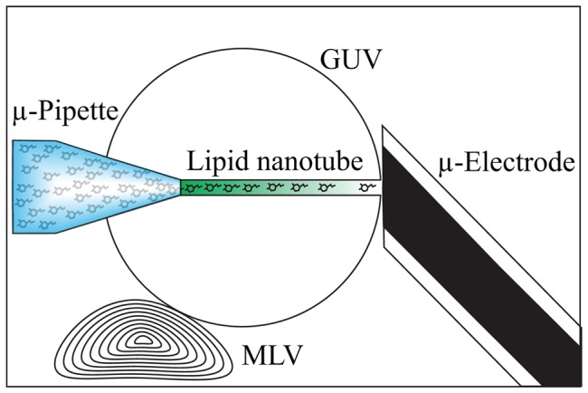
Figure 1. Experimental set up.

A giant unilamellar vesicle (GUV) is formed from a multilamellar vesicle (MLV) that is attached to the glass slide substrate. A micropipette filled with dopamine is inserted into the GUV and through the second membrane through electroporation. The pipette is then pulled back into the vesicle bringing with it a nanotube connecting the pipette to the outside of the vesicle. At the exit of the tube a micro electrode is positioned and the current through the nanotube is monitored with zero pressure applied through the micropipette.
